Written for Blind Low Vision NZ by Disha Rattu, Optometrist, Specsavers Albany, Auckland.
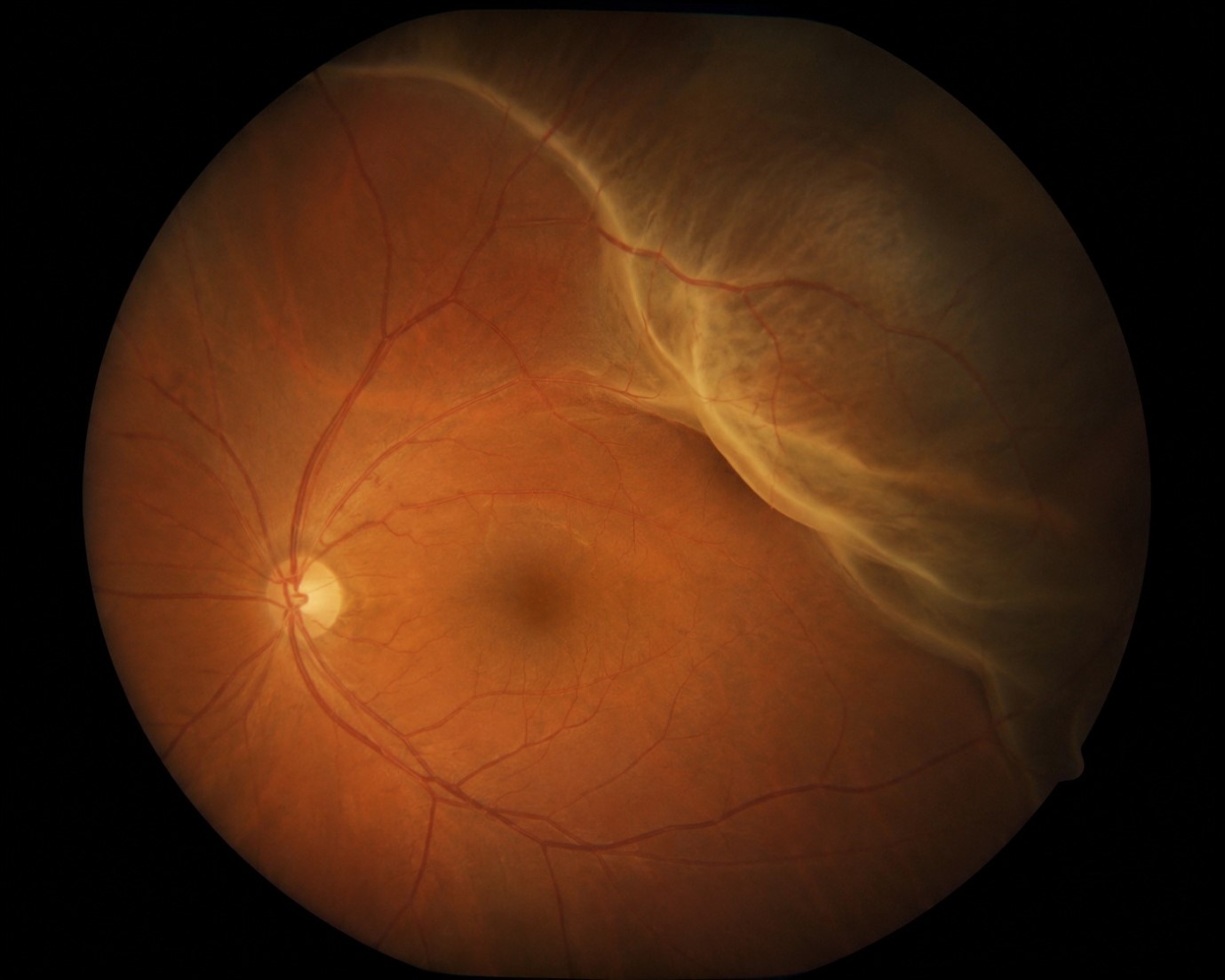
Image description: Representation of the inside of an eye with a partial retinal detachment.
What is retinal detachment?
In certain situations, the retina can form holes, tears or even completely pull away from its normal position and become a retina detachment. You can learn more about retinal detachment on the Specsavers website.
What is the retina?
The back of our eye is made up of three layers. The inner most layer is called the retina. Light will pass through the front of our eyes and onto the retina, which then transmits this information to the brain through our optic nerve, giving us the images we see in front of us. The most notable part of the retina is called the macula, which is responsible for our detailed central vision. The rest of the retina is responsible for our peripheral vision.
What are the symptoms of a retinal detachment?
Symptoms that can indicate a retinal detachment include:
- New Floaters in the eyes – floaters are tiny bits of protein deposits floating in the gel (called the vitreous) within the eye, and you can see them as squiggly lines, small dots, cobweb-like shapes, or rings. Most people have some floaters, however a sudden increase or change in floaters can be a warning sign of a retinal detachment.
- Flashes of Light in the vision
- A Dark Shadow, Curtain or Blurry patch obscuring part of the vision in the eye. This can be on the sides or in the centre
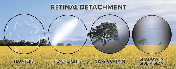
Image description: A slide titled “Retinal detachment” showing four circular images that represent common symptoms, including floaters shown as small shapes in vision, flashes of light shown as a bright area, a grey curtain effect shown as part of an image being covered by a grey area, and shadows or loss of side vision shown as a dark area on the side of an image. (Credit: Ophthalmology24)
Who is at risk of having a retinal detachment?
Anyone can develop a retinal detachment, but some factors that increase the likelihood include:
- Extreme near-sightedness (high myopia)
- Having a family member who had a retinal detachment
- Trauma or serious injury to the eye
- History of eye surgery, such as cataract surgery
- Age: A natural process related to ageing, called Posterior Vitreous Detachment (PVD), causes the jelly-like substance (vitreous) inside your eye to shrink away from the retina. Sometimes, the vitreous can pull on the retina and create retinal tears or a detachment.
Other eye conditions such as advanced diabetic retinopathy, inflammation in the eye, a tumour in the eye, or thin areas in your retina (such as lattice degeneration)
What should I do if I think I have a retinal detachment?
A retinal detachment is a medical emergency. If it is left untreated, more of the retina can detach and this increases the likelihood of permanent vision loss or blindness. A detached retina is an acute condition, which means the symptoms can come on quite suddenly. If you experience any of the symptoms of a retinal detachment, you should immediately contact your local optometrist, ophthalmologist or the nearest acute eye clinic.
How is a retinal detachment treated?
Urgent treatment is usually required to repair the retina and help to restore or prevent loss of vision. Treatment options vary depending on the severity and location of the retinal detachment.
Small tears in the retina, if detected early, may be treated by laser or cryotherapy (freezing) to seal the retina back down.
If the retina has become completely detached, there are three main surgical procedures that your ophthalmologist may recommend for you:
Vitrectomy – the vitreous gel in the eye is removed and, in turn, gas or oil is injected to help keep the retina in place to heal. After this, laser therapy is utilised to seal the retina in areas where there are tears or the retina is weak. You can watch a video explaining what a vitrectomy involves on the Thomas Eye Group YouTube channel.
Pneumatic retinopexy – laser or cryotherapy is utilised to reattach the retina to the wall of the eye. After this, a gas bubble is injected into the eye to hold the retina in place while it heals.
Scleral buckle – the retina is surgically reattached, after which a silicone strap is attached to the white part of the eye, helping the retina stay in place.
What can I expect during the recovery period?
There is an expected recovery period of about two to four weeks after a retinal detachment surgery. If the surgery involves a gas bubble or oil, you may be asked to position your head in a certain way (such as face down) for a time after surgery to ensure the bubble stays in place against the retina. You may be required to wear an eye shield while sleeping and use eye drops during this period. You will be asked to avoid strenuous activity (e.g. heavy lifting, sports) and keep the eye free of dust, dirt or water entering the eye until your eye has healed. If your surgery involved a gas bubble, you won’t be able to fly until your doctor says it’s OK to do so.
About the author:
Disha Rattu is in her third year of practicing as a Senior Optometrist based in Auckland, New Zealand. She currently practices at Specsavers Albany and holds a Bachelor of Optometry with Honours from the University of Auckland.

Image Description: Disha Rattu, optometrist, smiling and wearing a navy blue cardigan with a Specsavers logo on it.
Support and Empowerment
Blind Low Vision NZ provides various resources to assist people with sight loss. This includes mobility training with a white cane or guide dog, training on adaptive techniques and technology, and more! To learn about the support we can offer, call our friendly Contact Centre on 0800 24 33 33 or visit the Our Services page on our website.
References
Bogoev M.D., A. and Maria Bogoeva, M. (2025) Retinal detachment and warning signs you shouldn’t ignore, Ophthalmology24. Available at: https://www.ophthalmology24.com/retinal-detachment (Accessed: 08 May 2026).
Janelle (2023) How do you diagnose PVD or retinal tear, LVF Eye Centre. Available at: https://www.lvfeyecentre.org.au/how-do-you-diagnose-pvd-or-retinal-tear/ (Accessed: 08 May 2026).
Retinal detachment (no date) National Eye Institute. Available at: https://www.nei.nih.gov/eye-health-information/eye-conditions-and-diseases/retinal-detachment (Accessed: 08 May 2026).
Retinal Tears & Detachments (2024) Auckland Eye. Available at: https://www.aucklandeye.co.nz/eye-conditions/retinal-tears-detachments/ (Accessed: 08 May 2026).
Turbert, D. and Gregori, N.Z. (2023) Eye anatomy: Parts of the eye and how we see, American Academy of Ophthalmology. Available at: https://www.aao.org/eye-health/anatomy/parts-of-eye (Accessed: 08 May 2026).
Next :
Hmm, no more results were found, click here to return to all "News".